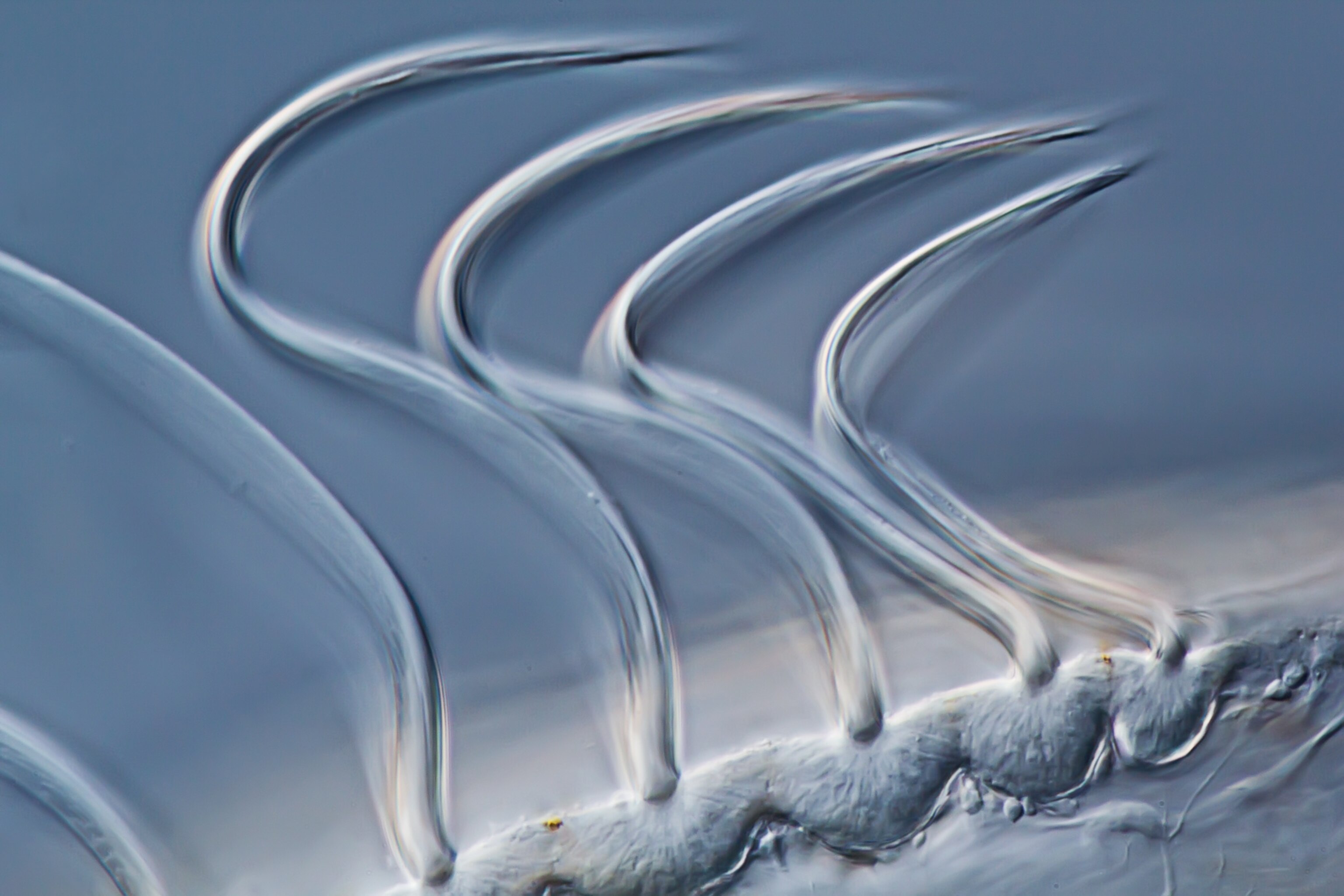
<p id="internal-source-marker_0.36668464934399725" dir="ltr"><strong>What looks like a tangle of Christmas lights is actually the <a href="http://science.nationalgeographic.com/science/health-and-human-body/human-body/brain-article/">brain</a> of a zebrafish embryo, winningly photographed for the <a href="http://www.nikonsmallworld.com/">2012 Small World Microphotography Competition</a>.</strong></p><p dir="ltr">Winners of the international contest were announced Tuesday. This shot—taken by Jennifer Peters and Michael Taylor of <a href="http://www.stjude.org/stjude/v/index.jsp?vgnextoid=f87d4c2a71fca210VgnVCM1000001e0215acRCRD">St. Jude Children's Research Hospital </a>and magnified 20 times—took first prize. It's believed to be the first image ever to show the formation of the blood-brain barrier in a live animal.</p><p dir="ltr">The barrier is a protective system of cells that filter the blood that flows to the brain. It allows nutrients and other necessities to pass but keeps out bacteria and other pollutants.</p><p dir="ltr">To create this shot, Peters and Taylor injected fluorescent proteins into a transparent zebrafish embryo. That let them see the brain's endothelial cells—which line the inner surface of blood and lymphatic vessels—and watch the development of a blood-brain barrier in real-time.</p><p dir="ltr">Their three-dimensional image was made with a confocal microscope, which colors blood vessels differently at different depths. (Confocal lenses capture light from a single plane of focus, rather than all available light.) Peters and Taylor stacked their colored images and compressed them into a single shot to show the complexity of the phenomenon they witnessed.</p><p dir="ltr">(See <a href="http://news.nationalgeographic.com/news/2011/10/pictures/111006-nikon-small-world-microphotography-competition-science/">pictures of 2011's best micro-photos</a>.)</p><p>—<em>Kate Andries</em></p>

1st Place: Bright Brain

What looks like a tangle of Christmas lights is actually the brain of a zebrafish embryo, winningly photographed for the 2012 Small World Microphotography Competition.

Winners of the international contest were announced Tuesday. This shot—taken by Jennifer Peters and Michael Taylor of St. Jude Children's Research Hospital and magnified 20 times—took first prize. It's believed to be the first image ever to show the formation of the blood-brain barrier in a live animal.

The barrier is a protective system of cells that filter the blood that flows to the brain. It allows nutrients and other necessities to pass but keeps out bacteria and other pollutants.

To create this shot, Peters and Taylor injected fluorescent proteins into a transparent zebrafish embryo. That let them see the brain's endothelial cells—which line the inner surface of blood and lymphatic vessels—and watch the development of a blood-brain barrier in real-time.

Their three-dimensional image was made with a confocal microscope, which colors blood vessels differently at different depths. (Confocal lenses capture light from a single plane of focus, rather than all available light.) Peters and Taylor stacked their colored images and compressed them into a single shot to show the complexity of the phenomenon they witnessed.

(See pictures of 2011's best micro-photos.)

Kate Andries

Image courtesy Jennifer Peters and Michael Taylor, Nikon Small World

Pictures: Best Micro-Photos of 2012

From baby spiderlings to a blood-brain barrier—see the best microphotos chosen in the annual Small World photo competition.

DON'T MISS THE REST OF THIS STORY!
Create a free account to continue and get unlimited access to hundreds of Nat Geo articles, plus newsletters.

Create your free account to continue reading

No credit card required. Unlimited access to free content.
Or get a Premium Subscription to access the best of Nat Geo - just $19
SUBSCRIBE

Read This Next

Is banning fishing bad for fishermen? Not in this marine reserve
SeaWorld allegedly violated the Animal Welfare Act. Why is it still open?
'World’s worst shipwreck' was bloodier than we thought

Go Further

Subscriber Exclusive Content

Why are people so dang obsessed with Mars?

How viruses shape our world

The era of greyhound racing in the U.S. is coming to an end

See how people have imagined life on Mars through history

See how NASA’s new Mars rover will explore the red planet

Why are people so dang obsessed with Mars?

How viruses shape our world

The era of greyhound racing in the U.S. is coming to an end

See how people have imagined life on Mars through history

See how NASA’s new Mars rover will explore the red planet

Why are people so dang obsessed with Mars?

How viruses shape our world

The era of greyhound racing in the U.S. is coming to an end

See how people have imagined life on Mars through history

See how NASA’s new Mars rover will explore the red planet